Identification of Bacteria Using Staining Techniques
In a simple stain, a bacterial smear is stained with a solution of a single dye that stains all cells the same color without differentiation of cell types or structures. The single dye used here is methylene blue, a basic stain. Basic stains, having a positive charge, bind strongly to negatively charged cell components such as bacterial nucleic.
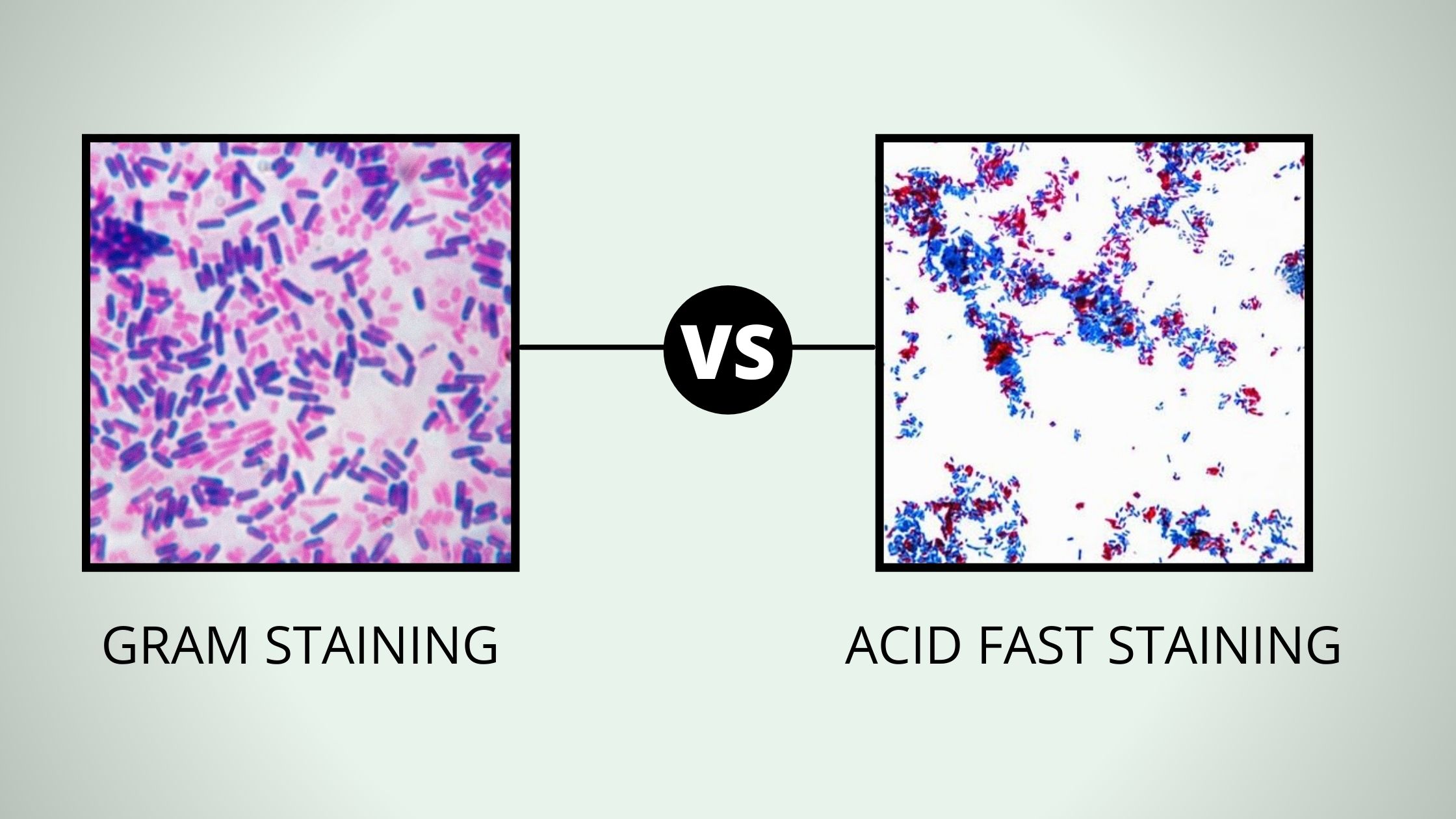
Comparison Between Gram Stain and Acid Fast
True to its name, the simple stain is a very simple staining procedure involving a single stain solution. Any basic dye, such as methylene blue, safranin, or crystal violet, can be used to color the bacterial cells. These stains will readily give up a hydroxide ion or accept a hydrogen ion, which leaves the stain positively charged.

Gram Staining Principle, Procedure and Results Learn Microbiology Online Medical laboratory
Staining is a technique where specimens are stained with specific dyes to create contrast between the specimen and its background.. Staining Methods - Simple Staining, Negative Staining, Gram's Staining and Acid-Fast Staining. In: Basic Techniques in Biochemistry, Microbiology and Molecular Biology. Springer Protocols.
Simple Staining Principle, Procedure, Results and Application Biology Ease
48 Share 1.6K views 2 years ago You will get to know the meaning of the terms 'staining' and 'stain' in the beginning of the video. Further, in this video you will find a detailed explanation.
Simple Staining Procedure, Principle, Result
A simple stain in microbiology is the addition of a cationic or positively charged dye to a slide. This dye then stains the clear or translucent cells on the slide or sample. By doing so, a.

Gram staining procedure Microbiology, Medical laboratory technician, Medical laboratory science
In a simple stain, a bacterial smear is stained with a solution of a single dye that stains all cells the same color without differentiation of cell types or structures. The single dye used here in our lab is methylene blue, a basic stain. Basic stains, having a positive charge, bind strongly to negatively charged cell components such as.
Simple Staining Procedure, Principle, Result
Grip the microscope slide in wood clip over a waste container bucket. Add methylene blue stain to the heat-fixed smear. There is no reason to cover the entire slide with stain. Just make sure to cover the smear with stain. Set a timer for 1.5 or 2 minutes. The stain will remain on the smear during this time.
Simple Staining Procedure. Download Scientific Diagram
Simple staining technique uses a single stain to visualize the bacteria, which produces a distinctive contrast between the organism and its background.

Simple staining Procedure and its Mechanism Microbiology with Sumi YouTube
Simple staining involves directly staining the bacterial cell with a positively charged dye in order to see bacterial detail, in contrast to negative staining where the bacteria remain unstained against a dark background.
Gram Staining Technique = Simple Explanation Via Animated Presentation (HINDI) By Solution
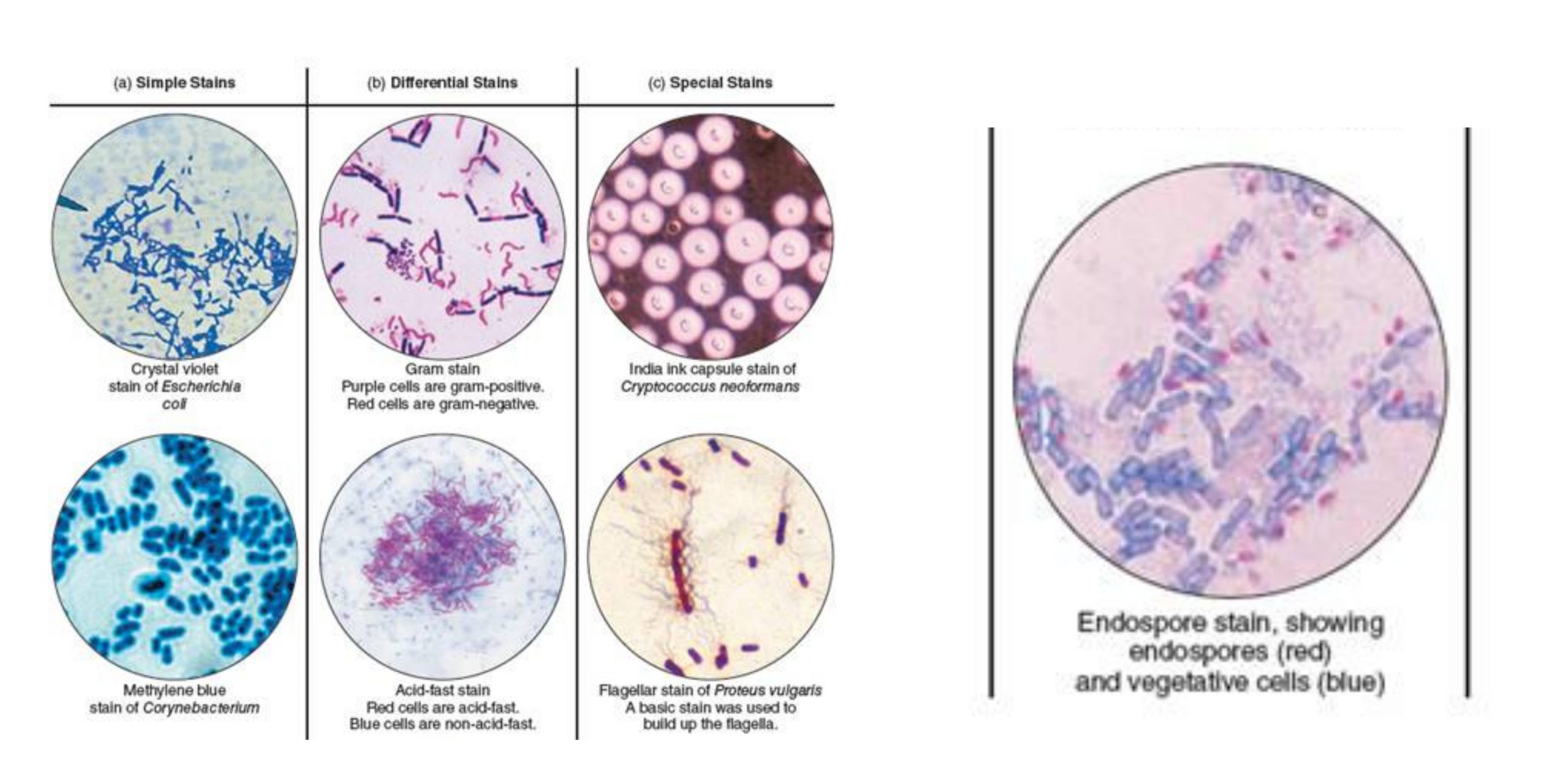
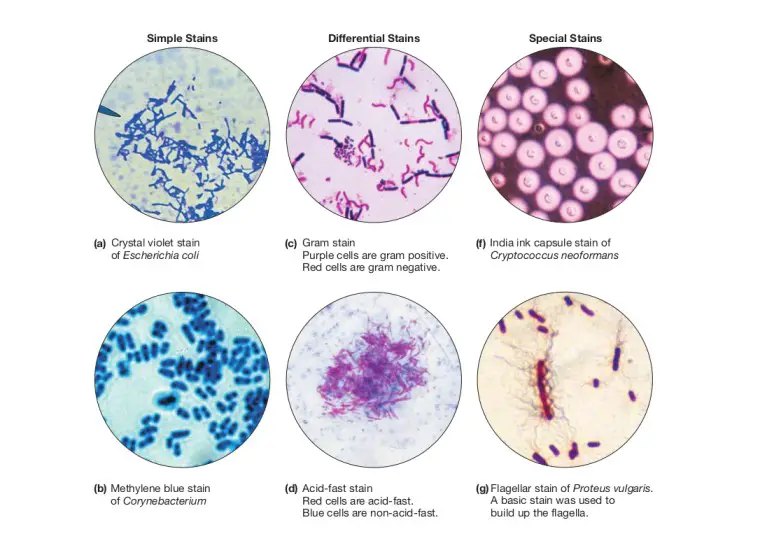
A simple stain will generally make all of the organisms in a sample appear to be the same color, even if the sample contains more than one type of organism. In contrast, differential staining distinguishes organisms based on their interactions with multiple stains. In other words, two organisms in a differentially stained sample may appear to.
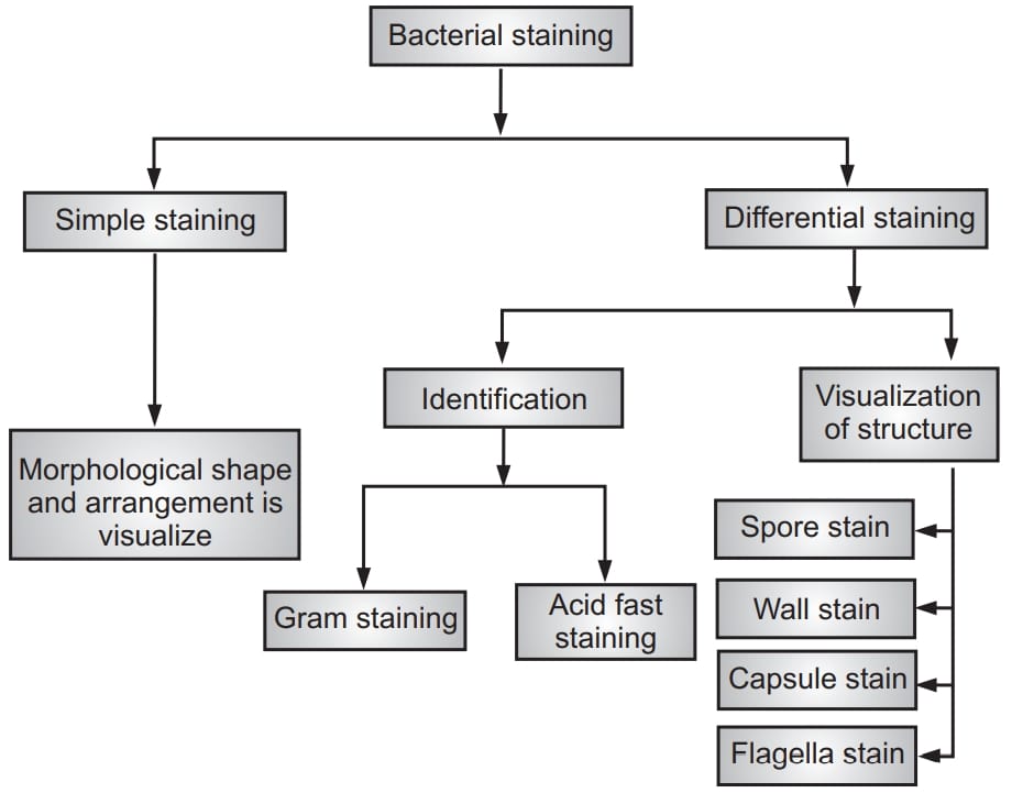
Types of Staining Techniques
Article Shared by ADVERTISEMENTS: The following points highlight the top five types of Staining. The types are: 1. Simple Staining 2. Differential Staining 3. Gram Staining 4. Acid Fast Staining 5. Endospore Staining. Staining Type # 1. Simple Staining: Colouration of microorganisms by applying single dye to a fixed smear is termed simple staining.

Microbial Staining= Simple Staining Gram Staining Acid Fast Staining Staining Technique
Article Joint by ADVERTISEMENTS: The following credits highlight to top five types away Staining. The types are: 1. Unsophisticated Dyeing 2. Differential Staining 3. Gram Staining 4. Acid Fast Staining 5. Endospore Dye. Staining Type # 1. Simple Staining: Colouration out human over applying unique dye to an fixed smear is termed easy staining.

Gram staining Principle, Requirements, Procedure and Microscopic Examination Online Science Notes
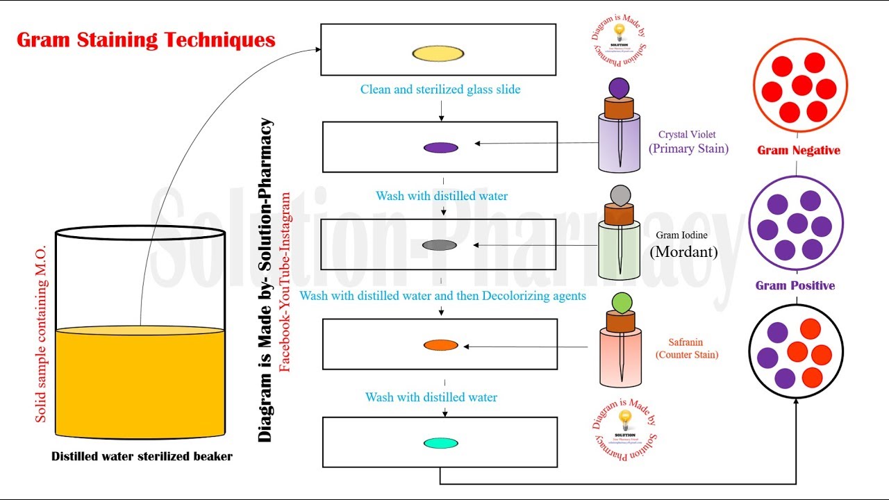
Grasp the slide with a slide holder and pass the smears through the upper part of a flame at least 2-3 times. Avoid overheating the slide. Place the slide on the metal stain rack over the sink. Cover the smears with the stain using the following times: Crystal violet: stain for 30 to 60 seconds.
Example in Lab in Which Simple Staining Would Be Used
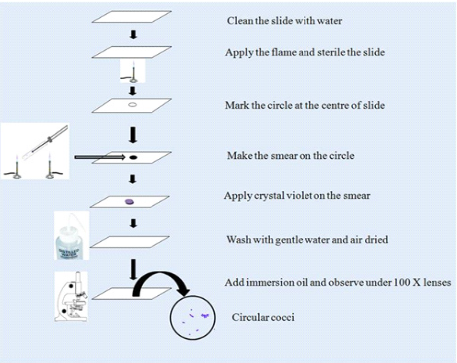
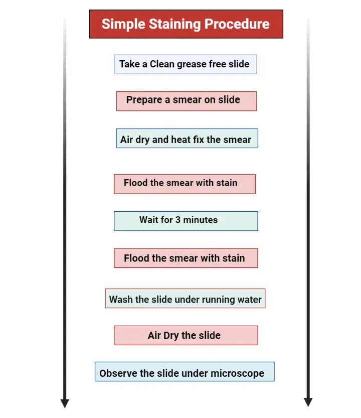
16: Simple Stain. Prepare a smear from a bacterial specimen. Prepare a simple stain of the smear. Use the microscope to identify features (shape, arrangement, size) of the bacterium. In order to stain the bacterial specimen for microscopy one must first prepare the smear on the slide. This basically involves three steps----transferring a liquid.
Microbiology Mania Gram Staining, Including Simple Staining Method {Lab 3 May 14, 2015}
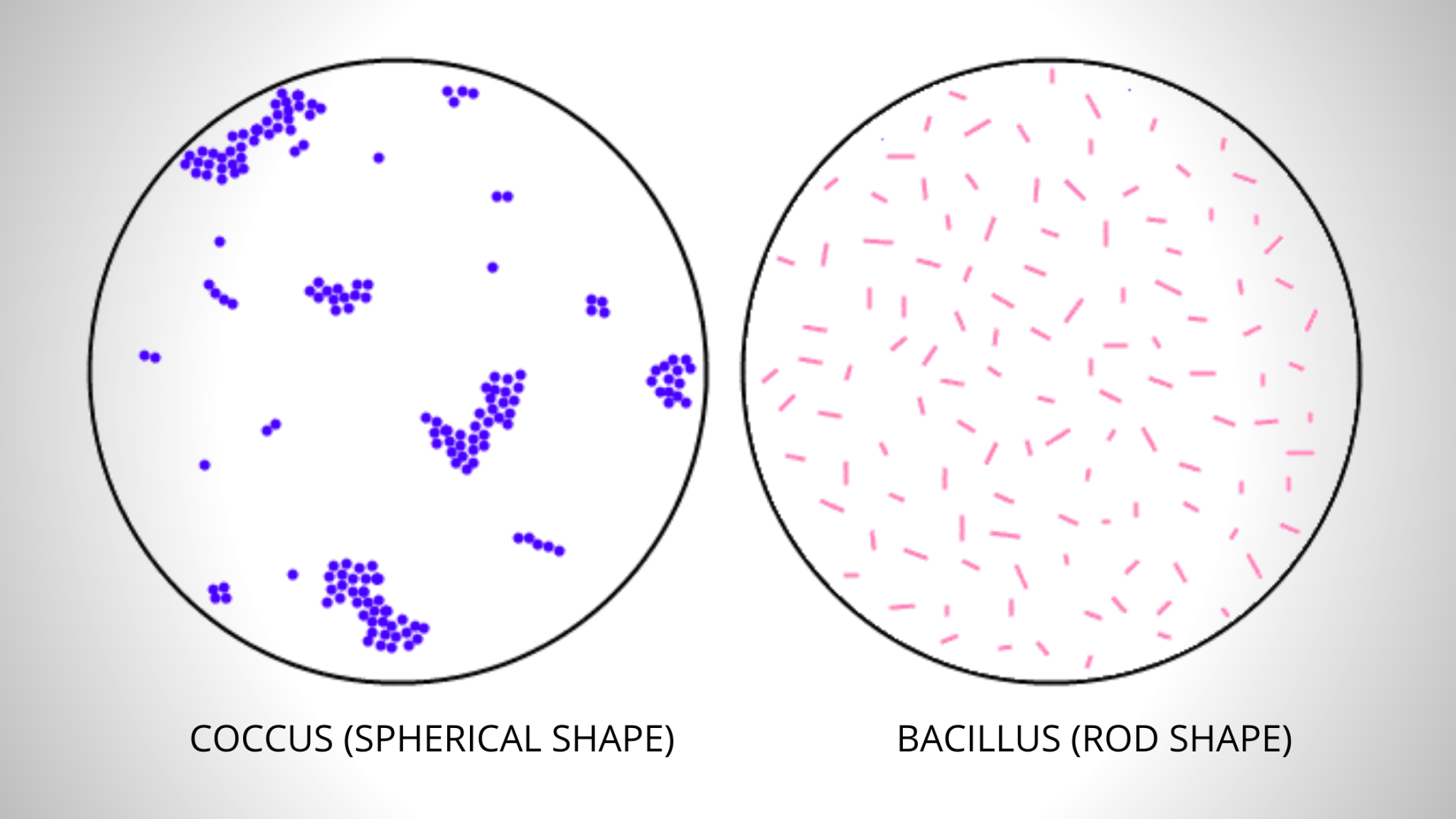
The purpose of simple staining is to elucidate the morphology and arrangement of bacterial cells. The most commonly used basic stains are methylene blue, crystal violet, and carbol fuchsin. Reagents and Equipment's for Simple Staining

Simple staining technique Online Biology Notes
Principle: Simple staining uses single basic dyes such as crystal violet which is dissolved in a solvent and applied to the microorganisms. The microorganisms give the colour characteristics of the staining solution. Because of which shape and size of microorganisms can be determined. Requirements: